Appointments Mon. to Fri. 8:30 a.m. to 6:00 p.m.
Talk to us!
(Call to national fixed network)

Mammography with 3D Tomosynthesis

Mammography with 3D Tomosynthesis is an advanced imaging exam for breast screening and diagnosis, allowing a more detailed assessment of breast tissue.

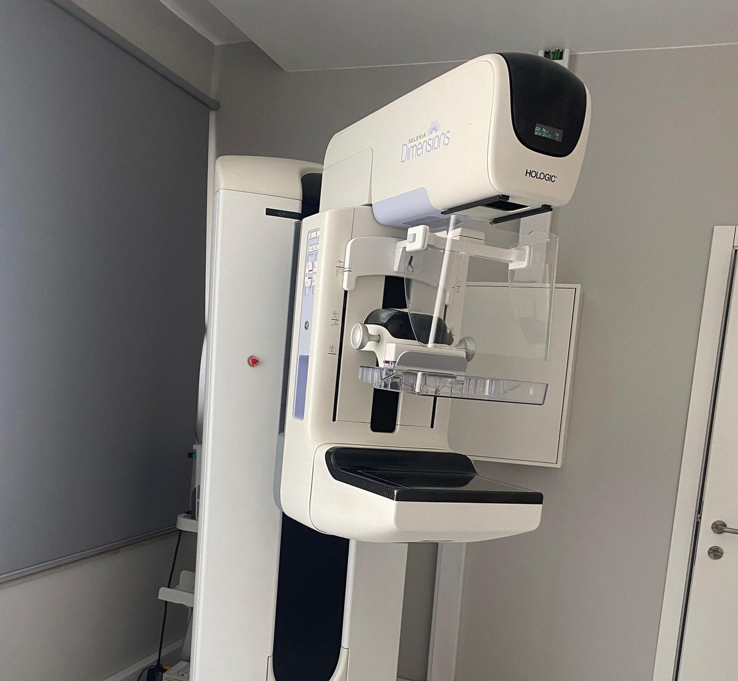
At NRD, in Lisbon, you can have your Mammography with 3D Tomosynthesis in a clinical setting specialised in diagnostic imaging, with technology dedicated to breast assessment.

What is it and what is it used for?

Mammography with 3D Tomosynthesis is an evolution of digital mammography. Like conventional mammography, it uses low-dose X-rays to study the breast, but allows a more precise analysis of breast tissue.

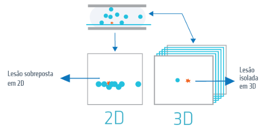
In traditional mammography, breast structures are represented in a two-dimensional image. This can cause different tissues to overlap, making it more difficult to see small lesions, masses or distortions.

With 3D tomosynthesis, the radiologist can assess the breast layer by layer, reducing this overlapping effect and obtaining a clearer reading.

This exam is used to support breast cancer screening and early diagnosis, as well as to clarify breast changes identified in previous exams, symptoms or situations of increased individual risk.

When breast cancer is detected at an early stage, before spreading to the lymph nodes or other parts of the body, treatment possibilities are generally more favourable.

What are the advantages of Mammography with 3D Tomosynthesis?

3D Mammography allows the radiologist to observe the breast with greater clarity, helping to better distinguish normal structures from changes that may require monitoring or further investigation.

A good way to understand this technology is to think of a book: if you only look at the cover, you cannot see all the pages. But when you open it, you can examine it page by page. 3D tomosynthesis follows the same principle, allowing breast tissue to be assessed layer by layer.

The main advantages of Mammography with 3D Tomosynthesis include:

  • better visualisation of masses, nodules, asymmetries and distortions;
  • greater assessment capacity in dense breasts;
  • reduction of doubts caused by tissue overlap;
  • support for the early diagnosis of breast cancer;
  • possibility of reducing the need for additional exams in some cases;
  • increased detection of invasive breast cancers, according to clinical studies referred to on the current page;
  • a more complete assessment when compared with two-dimensional mammography alone.

This technology represents an important advance in breast assessment, especially because it allows the identification of changes that could be less evident on a conventional image.


Differences between 2D mammography and 3D mammography


What is the price of Mammography with 3D Tomosynthesis?

The price of Mammography with 3D Tomosynthesis varies according to the health system, insurance or health plan you wish to use.

At NRD, the following agreements and prices are available for Mammography with Tomosynthesis:

  • ADSE: the patient only pays the co-payment, usually €16;
  • IASFA, ADM GNR, SAD PSP;
  • Insurers: Médis, Multicare, Advance Care, Allianz, Saúde Prime, Future HealthCare, SS CGD, SAMS Quadros, SAMS, Montepio, RNA, Generali, Mudum, MGEN, Aegon, Vitória, Una Seguros, other insurers;
  • Health plans: Medicare, Saúde Prime, Future HealthCare, Continente Wells, ACP, other health plans.

The final price should be confirmed when booking, according to the applicable agreement, insurance or health plan.

How is it performed?

Mammography with 3D Tomosynthesis is performed in a similar way to a conventional mammogram.

During the exam, the breast is positioned in the equipment and compressed for a few seconds. This compression is necessary to obtain clear images, reduce the radiation dose and improve diagnostic quality.

While the breast is compressed, the equipment acquires several images from different angles. These images are then reconstructed into thin slices, allowing a three-dimensional assessment of breast tissue.

The exam is quick. It may cause some discomfort due to compression, but this moment is brief. If you feel significant pain during the exam, you should inform the technician.

After the images are acquired, they are analysed by a radiologist, who prepares the corresponding report.


Mammography with 3D Tomosynthesis


What does it detect?

Mammography with 3D Tomosynthesis helps detect and characterise several breast changes, including:

  • breast nodules or masses;
  • microcalcifications;
  • asymmetries between the breasts;
  • architectural distortions;
  • changes suspicious for breast cancer;
  • lesions that may be hidden by tissue overlap;
  • benign changes that require monitoring or clarification;
  • changes that may require complementary exams, such as breast ultrasound, breast MRI or biopsy.

3D tomosynthesis has strengthened breast cancer screening, allowing the identification of changes that may be less evident on traditional mammography.

In which cases is it indicated?

Mammography with 3D Tomosynthesis may be indicated in several situations, including:

  • breast cancer screening;
  • breast assessment before the age range of organised screening, when clinically indicated due to symptoms, family history or increased individual risk;
  • study of dense breasts;
  • assessment of a nodule or palpable change;
  • localised breast pain;
  • nipple changes;
  • nipple discharge;
  • clarification of changes found in previous exams;
  • follow-up after breast surgery;
  • follow-up after oncological treatment;
  • personal or family history of breast cancer;
  • need for a more detailed assessment compared with conventional mammography.

The indication should always be considered according to age, clinical history, symptoms, family history and previous exams.

Preparation for Mammography with 3D Tomosynthesis

In most cases, Mammography with 3D Tomosynthesis does not require any special preparation. Even so, there are some important recommendations to ensure image quality and the correct clinical context for the exam.

Topic Guidance
Exam prescription You should bring the exam prescription, if applicable, especially if the exam is performed through an insurance, agreement or health subsystem.
Deodorant, creams or perfumes On the day of the exam, do not apply deodorant, cream, perfume, talcum powder or lotions to the breast or underarm area, as these products may interfere with the image.
Previous exams You should bring previous breast exams, such as mammograms, breast ultrasounds, MRIs or clinical reports. Comparison with previous exams is important for a more accurate assessment.
Pregnancy or suspected pregnancy Inform the team if you are pregnant or suspect you may be pregnant, so that the situation can be assessed before the exam is performed.
Breastfeeding If you are breastfeeding, you should inform the team when booking or before the exam.
Breast implants Inform the team if you have breast implants or have undergone breast surgery.
Clothing and accessories Comfortable clothing that is easy to remove from the waist up is recommended. You should remove necklaces or accessories that may interfere with the exam.
Breast tenderness If you still menstruate and have increased breast tenderness, it may be more comfortable to perform the exam in the days after your period.
Early arrival You should generally arrive a few minutes before the scheduled time to complete admission and exam preparation.
Fasting Fasting is not required for Mammography with 3D Tomosynthesis.

NRD will provide you with all the necessary instructions when booking.

When and how do you receive the results?

After Mammography with 3D Tomosynthesis is performed, the images are analysed by a radiologist, who prepares the corresponding report.

On the day of the exam, the team will inform you when and how you will be able to access your results.

Where to do it?

3D Mammography can be performed at NRD, at Avenida Columbano Bordalo Pinheiro, nº 11-B, ground floor, 1070-060 Lisbon (next to Praça de Espanha) or at X-Clinic, at Av. Eng. Duarte Pacheco, nº 26 – middle floor, 1070-110 Lisbon.

Does Mammography with 3D Tomosynthesis hurt?

The exam may cause some discomfort due to breast compression, but this compression only lasts a few seconds. If you feel significant pain, you should inform the technician during the exam.

Does Mammography with 3D Tomosynthesis involve radiation?

Yes. Like conventional mammography, it uses low-dose X-rays. The dose is controlled and appropriate for the diagnostic purpose.

Does 3D tomosynthesis replace conventional mammography?

3D tomosynthesis is an advanced form of mammography. It allows breast tissue to be assessed layer by layer and may offer greater detail than two-dimensional imaging alone.

What is the difference between 2D mammography and 3D mammography?

In 2D mammography, breast structures are observed in a flat image and may overlap. In 3D mammography, the breast is analysed layer by layer, which helps reduce this overlap and allows small details to be seen more clearly.

Is Mammography with 3D Tomosynthesis indicated for dense breasts?

Yes. It can be particularly useful in dense breasts because it helps reduce breast tissue overlap, which can make conventional mammography more difficult to interpret.

Can the exam detect breast cancer at an early stage?

Mammography with 3D Tomosynthesis is an important tool for breast cancer screening and early diagnosis. It helps identify suspicious changes that may require monitoring or further investigation.

How long does the exam take?

Image acquisition is quick, although the total time may vary depending on preparation, positioning and the possible need for additional images.

Do I need to bring previous exams?

Yes. Whenever possible, you should bring previous mammograms, breast ultrasounds, MRIs or reports. Comparing with previous exams is important to identify new changes or confirm the stability of already known findings.

Can I have the exam if I am pregnant or breastfeeding?

You should inform the team if you are pregnant, suspect you may be pregnant or are breastfeeding. In these cases, the need for the exam will be assessed according to the clinical reason and individual situation.