Spine magnetic resonance imaging (MRI) is a non-invasive, radiation-free imaging exam that allows a highly accurate assessment of the different regions of the spine: cervical, thoracic, lumbar and lumbosacral. It is frequently used in the diagnosis of disc herniations, nerve compressions, and inflammatory, traumatic or tumoral changes.
What is a Spine MRI?
Spine magnetic resonance imaging (MRI) is an exam that uses magnetic fields and radiofrequency waves to obtain detailed images of the structures of the spine. It allows clear visualisation of the intervertebral discs, spinal cord, nerve roots, joints and surrounding soft tissues.
Cervical Spine MRI
Cervical spine MRI assesses the first seven segments of the spine, which support the neck and protect the cervical spinal cord. It is indicated in cases of persistent neck pain, radiculopathy (pain radiating to the arms), neurological changes, suspected cervical disc herniation, trauma, assessment of spinal cord compression, among others.
Thoracic Spine MRI
Thoracic spine MRI examines the thoracic region, which corresponds to the twelve middle segments of the spine. It is especially useful in the investigation of thoracic back pain of unclear origin, tumours, infections, osteoporotic fractures and inflammatory diseases. It may also be indicated in the context of spinal cord assessment or in patients with systemic diseases that affect the spine.
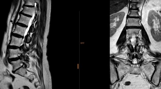
Lumbar Spine MRI
Lumbar spine MRI analyses the five lower segments of the lumbar spine, which support much of the body’s weight. It is frequently requested to investigate low back pain, disc herniations, nerve compressions causing sciatica, lumbar canal stenosis, degenerative changes, spondylolisthesis, post-traumatic lesions, etc.
Lumbosacral Spine MRI
Lumbosacral spine MRI focuses on the transition between the lumbar spine and the sacrum, including the sacroiliac joint. It is particularly relevant in cases of low back or sacral pain, suspected sacroiliitis, ankylosing spondylitis, inflammation of the sacroiliac joints, or when the aim is to study the origin of neurological symptoms in the legs associated with the lowest region of the spine.
What is the price of a Spine MRI?
The price of a spine MRI is 275€, when performed on a private basis. However, it can also be performed through several agreements, including:
- ADSE: If the spine MRI is performed under ADSE, the patient only pays a fee of 30€;
- IASFA, ADM GNR, SAD PSP;
- Insurance companies: Médis, Multicare, Advance Care, Allianz, Saúde Prime, Future HealthCare, SS CGD, SAMS Quadros, SAMS, Montepio, RNA, Generali, Mudum, MGEN, Aegon, Vitória, Una Seguros, other insurers.
- Health plans: Medicare, Saúde Prime, Future HealthCare, Continente Wells, ACP, other health plans.
- Protocols: NRD has protocols with several clinics and institutions that allow you to undergo the exam at very competitive prices. For a spine MRI, the price through protocol is 220€.
The price varies depending on the healthcare system or insurance you wish to use.
Find out more about the price of MRI exams.
How is it performed?
The patient is positioned lying on a table, with the spine supported on a coil appropriate to the region being studied. The head may be positioned in a specific coil in the case of a cervical spine MRI. Images are acquired in several planes, with thin, high-resolution slices.
The MRI scanner is open at both ends.
In some cases, it may be necessary to administer intravenous contrast, especially to assess infections, tumours or inflammatory lesions.
During the exam, it is essential that the patient remains still. Ear protection is provided due to the noise generated by the scanner. The exam may take between 30 and 50 minutes, depending on the region(s) being assessed and whether contrast is required.

What does it detect?
Spine MRI allows the detection of changes such as:
- Disc herniations and protrusions;
- Stenosis of the spinal canal or neural foramina;
- Lesions of the spinal cord and nerve roots;
- Bone, spinal cord or meningeal tumours;
- Inflammatory conditions (spondylodiscitis, ankylosing spondylitis);
- Fractures due to osteoporosis or trauma;
- Spondylolisthesis and degenerative changes (facet osteoarthritis, disc disease);
- Congenital malformations of the spine;
- Among others.
In which cases is it indicated?
Your doctor may request a spine MRI in situations such as:
- Persistent pain in the cervical, thoracic or lumbar spine;
- Suspected disc herniation with nerve root compression;
- Neurological symptoms such as tingling, numbness or weakness;
- Investigation of spinal stenosis or myelopathy;
- Suspected vertebral infection or tumour;
- Post-trauma or post-surgical assessment;
- Monitoring of inflammatory diseases;
- Among others.
Preparation and guidelines for a Spine MRI
Before undergoing an MRI at NRD, please consider the following:
| Topic | Guidance |
| Exam prescription (paper copy) | You must bring the exam prescription in paper form. |
| Fasting | Most MRI exams do not require fasting. In cases with contrast, fasting for 3 hours is usually necessary (confirmed when booking). |
| Early arrival | Arrive 15 minutes before the scheduled time. |
| Medication | You may take essential and unavoidable medication with a minimum amount of water. |
| Metal objects | Remove all metal objects (e.g. earrings, bracelets, necklaces, piercings, hairpins, removable prostheses, etc.). |
| Pregnancy | Inform the NRD healthcare professional if you are pregnant. |
| Exams / Tests / Info | Bring previous imaging exams, relevant clinical information and recent laboratory tests. |
| Glucose monitoring implant | Do not place the implant, as it will have to be removed before the exam. |
| Make-up | Do not wear make-up, as some products may affect the exam. |
| Valve and/or orthopaedic prostheses | If you have valve and/or orthopaedic prostheses, bring proof describing and specifying the material composition. |
| Hearing aids | Inform us if you use a hearing aid, as it must be removed before the exam. |
| Dental prostheses | Inform us if you use a dental prosthesis, as it must be removed before the exam. Also inform us if you have dental implants. |
| Orthodontic appliances | Inform us if you wear orthodontic appliances. A test may be necessary to assess compatibility with MRI. |
| Pacemakers and implantable devices | NRD does not perform MRI exams on people with pacemakers or implantable devices (e.g. cochlear implant), and it is recommended that the exam be carried out in a hospital setting. |
Throughout the MRI exam, you will be continuously monitored by the NRD imaging technologist responsible for your exam. An alert system will also be at your disposal, which you can activate if needed.
You should remain still during the exam to ensure high-quality images.
The entire NRD team will be at your disposal to answer any questions that may arise.
After the exam, you can return to your usual daily activities.
When and how do you receive the results?
You will receive the MRI results within 7 days after the exam, by email or in paper form at the clinic. On the day of the exam, you will take a CD with the images with you.
Where can you have it done?
The MRI can be performed at NRD, at the following address:
Avenida Columbano Bordalo Pinheiro, nº 11-B, r/c, 1070 – 060 Lisbon (near Praça de Espanha)